Illustrations of the Liver Sinew Channel
It's said that a picture is worth a thousand words. As an educator and anatomist, I find this is especially true. I have often found that very complex anatomical relationships can be understood much more easily by a very good illustration or image. Cadaver dissection is probably the best way to appreciate these relationships, but here are several images that are most certainly better than 'a thousand words and can help until you can get to a dissection.
Note: we are working on scheduling a 3 day cadaver lab on the East Coast with Sports Medicine Acupuncture and I will be doing a 1 day in Miami in September 2023 with Michael Corradino. Also note that I will be speaking on the QL and the LIV jingjin at the upcoming FSOMA conference in August 2023
Now on to the anatomy!
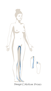
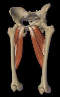
Classically, the Liver sinew channel terminates at the groin. Above is a wonderful illustration from Jennifer Black at Holism Prints which shows the classical view described in Ch 13 of the Lingshu. Below is my anatomical interpretation of this portion of the channel at the groin which involves the anterior adductors (adductor longus, brevis, pectineus, gracilis). All attach to the pelvis on it's inferior surface. This matches the classical view, but does this sinew channel end there?
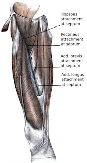
To the left is an image from Toldt's Atlas of Human Anatomy. The patella is facing to the left in this illustration. The perspective is anteromedial which gives a great view of the region of the Liver sinew channel. I produced the labels of the adductor attachments in the intermuscular septum, sometimes referred to as the subsartorial canal (it is deep to the sartorius muscle) and also it is known as Hunter's canal. I teach some manual techniques to open this space and also needle from it angled either anterior into trigger points of the quadriceps or posterior into the adductors. There are blood vessels and nerves running in this space and I feel this canal is where the Liver primary channel runs.
I love this image since is depicts how this myofascial plane continues beyond the groin and into the anterior portion of the pelvis. This entire atlas is amazing!
Toldt's Atlas is from the early 1900s. Let's go back a bit further to 18th century to view an image from Bernhard Siegfried Albinus, a German anatomist, who guided engraver Jan Wandelaar to make a series of fantastic engravings portraying different layers of the body. Here is the deepest layer which beautifully depicts the liver sinew channel all the way up through the anterior pelvis and into the spinal column to the diaphragm!

Check out my video that covers this sinew channel from the toes all the way to the diaphragm. Make sure to like the video and let me know if you have any thoughts, questions or considerations in the comment section.


No comments:
Post a Comment